Our annual Halloween event, It Came From the Archives, will not be happening this year for obvious reasons. For the past six years, we have enjoyed sharing a variety of materials from our collections in a casual open house setting in the library. 

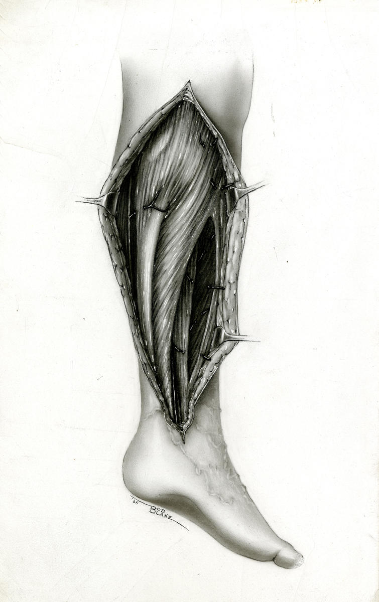
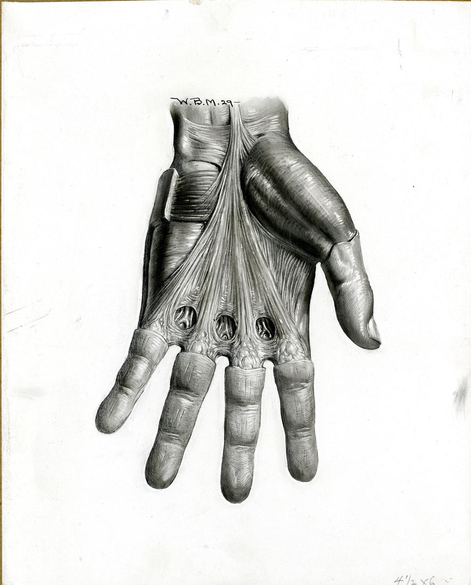
While we try to select different items each year for display, some of our favorite things to share every year are medical illustrations. Duke University Medical Center was among the first educational institutions in the United States to provide medical illustration services. Artwork was created with traditional and digital media and includes surgical and anatomic drawings, schematic and mechanical drawings, and a variety of designs. The images produced appear in books, journals, patient education materials, presentation materials, web sites, and occasionally on the pages of some of the nation's more prominent newspapers and covers of scientific journals. 

Medical illustration was present at Duke from the beginning. Max Brodel, the father of medical illustration from Johns Hopkins, sent his daughter, Elizabeth, to Duke in 1930 upon the opening of the school. In 1935, Elon H. Clark was appointed director of the Department of Medical Art and Illustration. As the director, Clark led a service-oriented research program that concentrated on the development of improved cosmetic prostheses, particularly facial restorations. Medical Illustration staff was also responsible for the coordination of pamphlets, charts, illustrations, and photographs used in many Duke University and Duke University Medical Center publications. 

In 1949, Robert Blake became part of the Duke faculty, and finally Coordinator of Medical Art (circa 1971) and Acting Director of Audiovisual Education, following Elon Clark's retirement. The Department of Medical Art and Illustration became the Division of Audiovisual Education in 1966, and consisted of three branches: medical art, medical photography, medical films and medical television. In 1997, Audiovisual Education became the Division of Educational Media Services (EMS), and consisted of four branches: medical photography, medical art, instructional television, and media coordination. The division was dissolved in 2005. 

In a world with abundant access to photography and audiovisual recordings, it might be hard to imagine a time when illustration was a vital part of medicine. These drawings are both educational and undeniable works of art. This post includes some of our favorites from the Robert L. Blake Papers and the J. Deryl Hart Papers and Records. We think that these illustrations are great representations of the talent of the artists, the educational value of their diagrams, and even a glimpse that Blake’s sense of humor! You may also view more of these illustrations on MedSpace.

